
Case Reveals Sarcoidosis Diagnosed with Endoscopic Ultrasound Elastography


Researchers at the University of Texas Health Science Center, in Houston, have described a sarcoidosis diagnosis by endoscopic ultrasound elastography.
But the report, published in the journal Endoscopic Ultrasound under the title “Endoscopic ultrasound elastography to diagnose sarcoidosis,” suggests that more studies are needed before the method can be used as a routine diagnostic tool for sarcoidosis.
The patient, a 54-year-old man, sought care after experiencing pain in the lower parts of his stomach during the past three months. The man was often nauseous, and had lost almost 14 kg (30 lbs.) from appetite loss.
Laboratory tests showed the man had anemia and abnormally high blood calcium levels. During an ultrasound, doctors noted a lump of tissue next to the pancreas, which prompted them to continue the investigation using computed tomography (CT) scans.
The scans revealed lymphadenopathy, or abnormal lymph nodes, around the liver and pancreas. The man’s ACE (angiotensin converting enzyme) showed high levels of the enzyme along with increased levels of antibodies in his blood. About 60 percent of sarcoidosis patients have high levels of the enzyme, which is believed to be produced by the immune cells forming sarcoidosis granulomas.
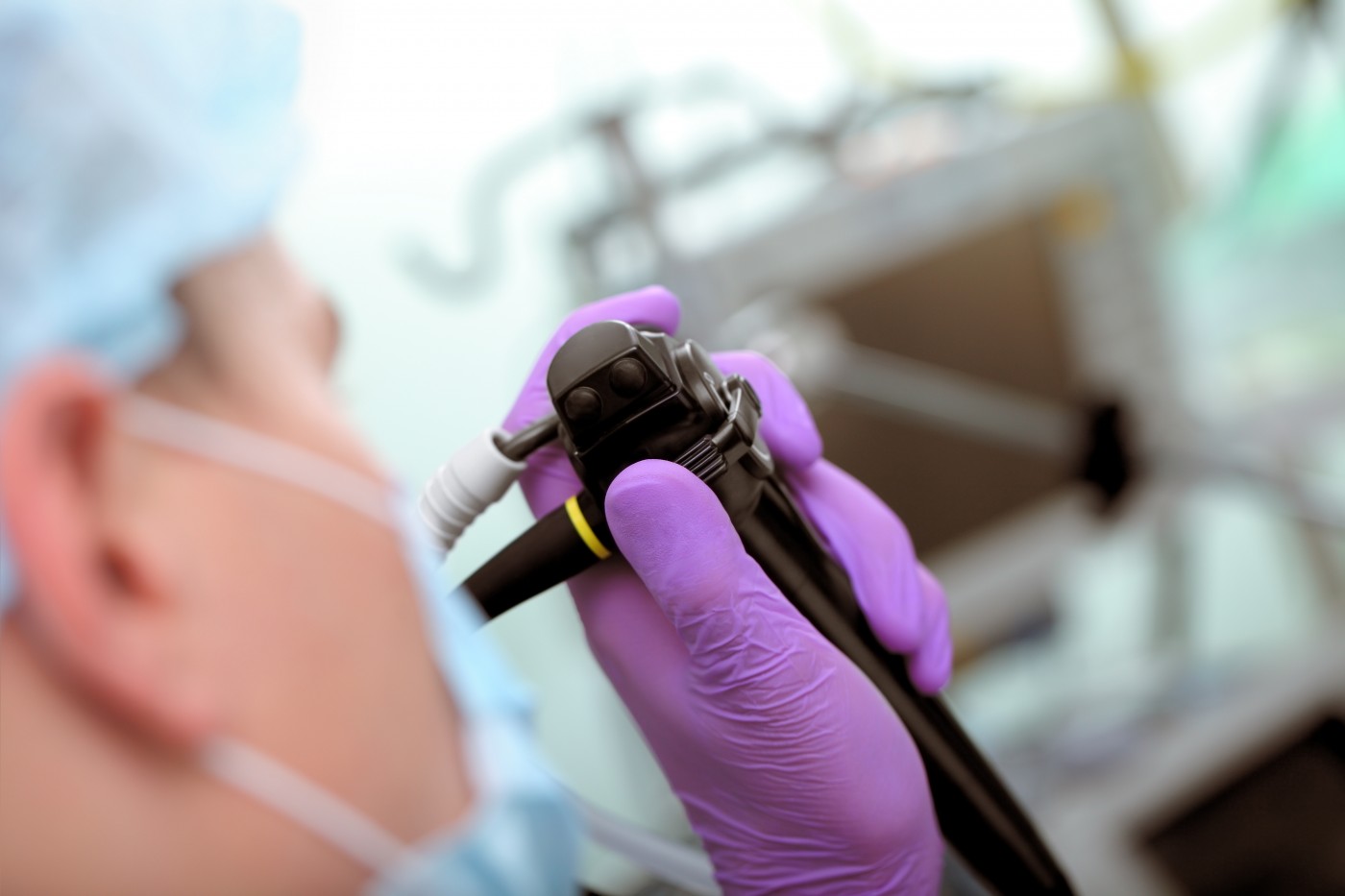
An endoscopic ultrasound combined with elastography showed several large lymph nodes next to the pancreas and liver. Elastography allows physicians to see the difference between harder tissues, most often being malignant tumors; and softer, more elastic tissues indicating other types of disease.
Biopsies by aspiration of fluid from the enlarged lymph nodes, showed no signs of infection but indicated the presence of granulomas, the hallmark of sarcoidosis, which together with the pattern of enlarged lymph nodes and laboratory findings resulted in the sarcoidosis diagnosis.
While elastography could be a promising tool for sarcoidosis identification, other elastography patterns in patients with sarcoidosis have been reported.