
Ground Glass Opacity a Rare Manifestation of Sarcoidosis, According to Chinese Study


Sarcoidosis is often detected through the identification of specific characteristics on high-resolution computed tomography (HRCT). But some rare manifestations of the disease may easily lead to misdiagnosis for other conditions.
Researchers at the First Affiliated Hospital of Dalian Medical University in China recently presented the case of a patient who exhibited an atypical manifestation of sarcoidosis called ground glass opacity that may also be indicative of lung diseases.
Their study, “Predominant diffuse ground glass opacity in both lung fields: A case of sarcoidosis with atypical CT findings,” was published in the journal Respiratory Medicine Case Reports.
Ground glass opacity is a nonspecific finding on HRCT scans that may be a consequence of thickening of the alveolar walls or septal interstitium, partial filling up of the lung alveoli, or a combination of both. Ground glass opacity may be indicative of a number of diseases, including tuberculosis, nonspecific interstitial pneumonia, pulmonary alveolar proteinosis, alveolar hemorrhage, and sarcoidosis.
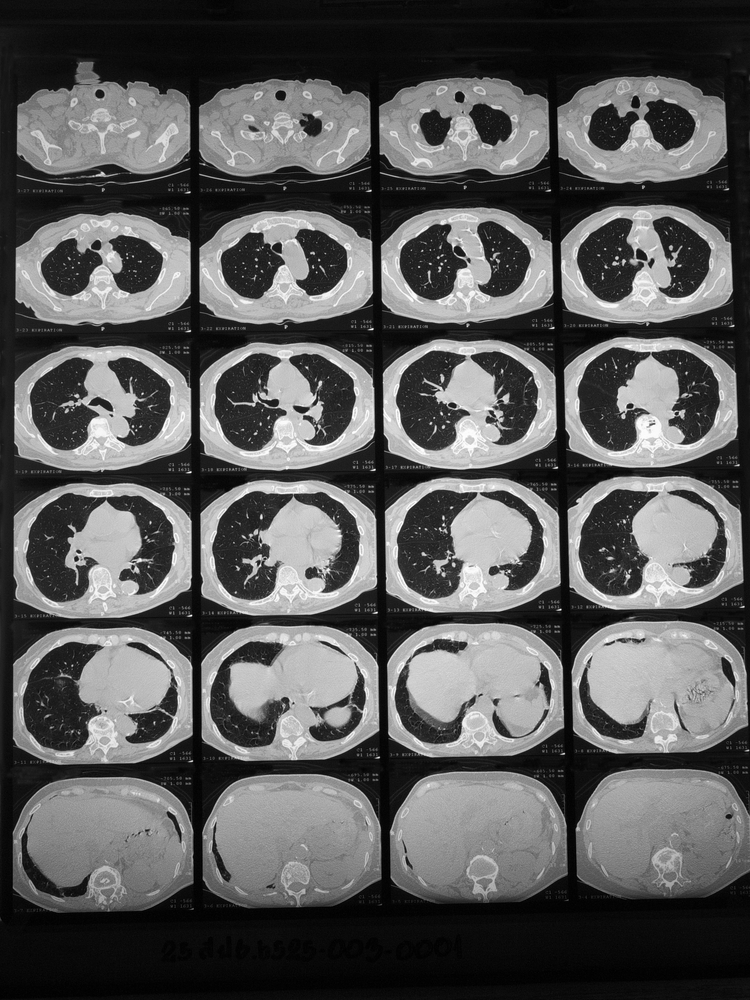
Now, the researchers presented the case of a 40-year-old Chinese woman who came to the hospital with a cough and recurrent skin rash. HRCT revealed large-scale ground glass opacity and minor lymphadenopathy.
The patient did not have a fever, which led the physicians to exclude tuberculosis. The presence of hilar lymphadenopathies usually indicates sarcoidosis, but the woman had only minor lymphadenopathy, which is not a common sarcoidosis manifestation.
After performing a battery of tests, researchers still could not exclude lung cancer, nonspecific interstitial pneumonia, alveolar hemorrhage, or pulmonary alveolar proteinosis. The patient underwent bronchoscopy to confirm intrathoracic sarcoidosis, but the small sample size and lack of clinicoradiological evidence prevented the diagnosis.
To obtain a large tissue mass and confirm diagnosis, a thoracoscopy was performed. No cancer cells were present, the findings were not typical for pulmonary alveolar proteinosis or alveolar hemorrhage, and the granulomas were not characteristic of nonspecific interstitial pneumonia, which together led to the eventual diagnosis of sarcoidosis.
“Predominant ground glass opacity in both lungs is a rare finding in intrathoracic sarcoidosis, as ground glass opacity is a nonspecific sign of lung sarcoidosis,” the authors said in their study. “In the absence of clinicoradiologic evidence to support the diagnosis of sarcoidosis, the diagnosis needs to be confirmed based on pathological evidence.”