
Thoracic Sarcoidosis Easier to Diagnose Using High-resolution CT, Study Says


Use of high-resolution computed tomography (HRCT) imaging technique can help identify features of thoracic sarcoidosis, researchers in India report. Because of its superior imaging capacity, they said, this method may allow clinicians to identify subtle chest abnormalities commonly undetectable by standard methods.
The results were published in the study “Thoracic Sarcoidosis: Imaging with High Resolution Computed Tomography” in the Journal of Clinical and Diagnostic Research.
Sarcoidosis is a multi-organ disease of unknown origin. It commonly affects patients’ lungs and is characterized by granulomas, which are accumulations of immune cells associated with increased inflammation, but also alterations of their thoracic lymph nodes, which in severe cases can be fatal.
Diagnosis can be difficult, requiring thorough radiological examination and lung function tests. In many cases, lung tissue biopsies are required to confirm the presence of the granulomas.
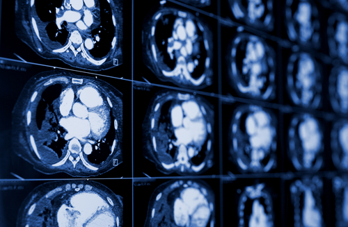
Imaging techniques are essential for the diagnosis, but also are used in follow-up of sarcoidosis patients. Standard chest radiography is the most common imaging method used. But it has detection limitations because of its low resolution. Computed tomography (CT) has a better imaging resolution, making it a more sensitive method to detect tissue and lymph node abnormalities.
Researchers evaluated the features of thoracic involvement in 40 patients with confirmed sarcoidosis using high-resolution CT, or HRCT, imaging.
HRCT had a superior capacity to detect disease features compared to standard chest radiography, the study’s authors found. HCRT could not only detect the pattern of the lesions and their distribution, but also could allow a more accurate assessment of the stage and activity of the disease. In addition, HRCT could detect abnormal, subtle tissue lesions that were not detected by conventional imaging.
“HRCT allows differentiation between active inflammation representing reversible disease from irreversible lung disease and fibrosis. Thus, it helps in prognosticating the disease and in guiding therapy,” the authors wrote.
“HRCT utilizes thin sections and high frequency reconstruction algorithms to generate images resulting in better characterization and definition of the abnormalities of the lung parenchyma [tissue],” they concluded. “HRCT is superior to conventional CT for assessing subtle parenchymal lesions and helps to differentiate active lesions from end stage disease.”