Researchers Propose Full-Body Scans for Cardiac Sarcoidosis Patients to Identify Secondary Disease Sites
Written by |

Patients with suspected cardiac sarcoidosis should be monitored for potential sarcoidosis lesions in other organs besides the heart, according to researchers at the University of Illinois at Chicago (UIC), who found that 40% of examined patients had sarcoidosis in other areas of the body.
The study, “FDG PET-CT findings of extra-thoracic sarcoid are associated with cardiac sarcoid: A rationale for using FGD PET-CT for cardiac sarcoid evaluation,” also demonstrated that a new imaging method, where patients maintain a high-fat, low-sugar diet for 72 hours before the scan, is better at detecting lesions than older methods.
The study was published in the Journal of Nuclear Cardiology.
“This study indicates that more extensive, full-body scans of patients with known cardiac sarcoidosis can uncover secondary sites of the disease in a significant number of patients,” Nadera Sweiss, professor of rheumatology at the UIC College of Medicine and a study co-author, said in a news release by Jacqueline Carey.
“Knowing there is disease in organs other than the heart changes the way we approach treatment — it allows us to more accurately stage the disease and treat it accordingly,” Sweiss added.
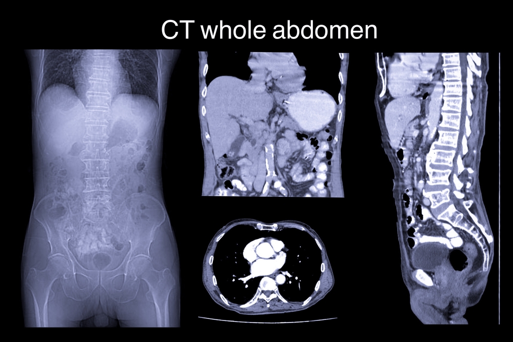
When doctors suspect sarcoidosis, they commonly use positron emission tomography (PET) and computed tomography (CT) scans to aid in diagnosis, including a combination of the two. However, the images are often unclear when looking for a diagnosis. This leads to the risk of failing to detect a sarcoidosis lesion, which can be common, Sweiss said.
To improve imaging results, researchers recently developed a protocol in which patients eat a high-fat, low-sugar diet for 72 hours before the scans. Previous protocols required a 24-hour diet.
Studies have shown that the new method is better at diagnosing cardiac sarcoidosis. Following this discovery, Sweiss and colleagues set out to test if the presence of cardiac sarcoidosis could indicate the presence of other lesions.
“We wanted to know if patients being evaluated for cardiac sarcoidosis would benefit from more thorough imaging of the body beyond the usual torso scan, using this new technique,” Sweiss said.
Scanning 188 patients with known sarcoidosis, from their nose to their knees, allowed the team to identify 20 patients with cardiac lesions. Among them, 40% also had lesions in other locations.
Based on the results, researchers proposed that a high-fat, low-sugar diet for 72 hours combined with full-body PET/CT imaging should be considered when assessing patients with cardiac sarcoidosis. Nonetheless, the team also emphasized that this diagnostic technique needs to be further tested.


