MRI Screenings Show Spinal Sarcoidois More Common Than Previously Thought, Study Reports

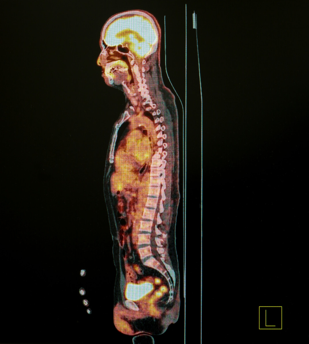
Highly sensitive magnetic resonance imaging (MRI) of the spinal cord shows that spine involvement is prevalent in sarcoidosis patients, mostly affecting the cervical region, a detailed re-analysis of imaging data reports.
The study, “Imaging findings in spinal sarcoidosis: a report of 18 cases and review of the current literature,” was published in The Neuroradiology Journal.
Spine involvement in sarcoidosis is considered to be a rare event due to a lack of tools that can differentiate between spinal manifestations in sarcoidosis and those observed in other neurological conditions affecting the spine.
The researchers in this study hypothesized that the prevalence of spinal sarcoidosis is higher than what has previously been reported. To examine this theory, they re-assessed the available MRI spinal imaging data for 18 cases of neurosarcoidosis to better understand the spinal deficits in these cases and patient response to treatment.
Analyzed cases spanned a 15-year period, and were shortlisted from the archives of the University of Iowa Hospitals and Clinics in the U.S., based on the availability of spine MRI results. The age of the patients ranged from 31-70 years at the time of spinal manifestation.
Of these 18 cases, MRI imaging data revealed some form of spinal abnormality in 72% (13 cases) of the patients analyzed. The cervical region of the spinal cord was affected in eight patients, more than what was found for the thoracic region, which was affected in six patients. Only four cases reported the involvement of the entire spinal cord.
The protective tissue (meninges) surrounding the skull and spinal cord consists of three layers: dura, arachnoid, and pia matter. Dura is the outermost layer, while pia matter is the innermost one. Researchers noted an enhanced dura layer (leptomeningeal enhancement) in eight (61%) of the cases, while a thick pia matter (pachymeningeal enhancement) was observed in three (23%) patients.
Using diffusion-weighted MRI images, lesions were detected in the spinal cord (intramedullary enhanced lesion) in six cases (38%), and the average length of the lesion was 4.3 spinal segments (the length of the spinal column consists of 31 segments).
Additionally, biopsy results confirmed the bone in the vertebral column was affected in two patients (15%).
Brain MRI is frequently used in patients with sarcoidosis. In the study, brain pachymeningeal abnormality was observed in 10 cases, while brain MRI from one patient showed the skull bone was affected.
All patients had received corticosteroids initially, followed by other immunosuppressivetreatment if necessary. Follow-up data were available for 93%, or 12 of the patients, with a mean follow-up period of 23.2 months.
At follow-up, imaging results indicating a complete or near complete reversal of the dura and pia matter thickening was observed in eight patients (66%), with mean improvement onset at 14 months after treatment.
Partial imaging improvement indicating moderate alleviation of spinal cord symptoms was reported in three patients (25%).
“Spinal sarcoidosis was previously considered uncommon but is being increasingly recognized with widespread use of magnetic resonance imaging,” the researchers wrote.
Based on the results, the team suggested that “these findings are helpful to distinguish SS [spinal sarcoidosis] from other neurological disease primarily affecting the spinal cord.” The team also believes that diffusion-weighted MRI images may offer insights about “disease activity and parenchymal injury” in sarcoidosis patients.


