
First Total-body PET Scanner Promises to Increase Sensitivity of Diagnosis and Treatment of Sarcoidosis, Other Diseases
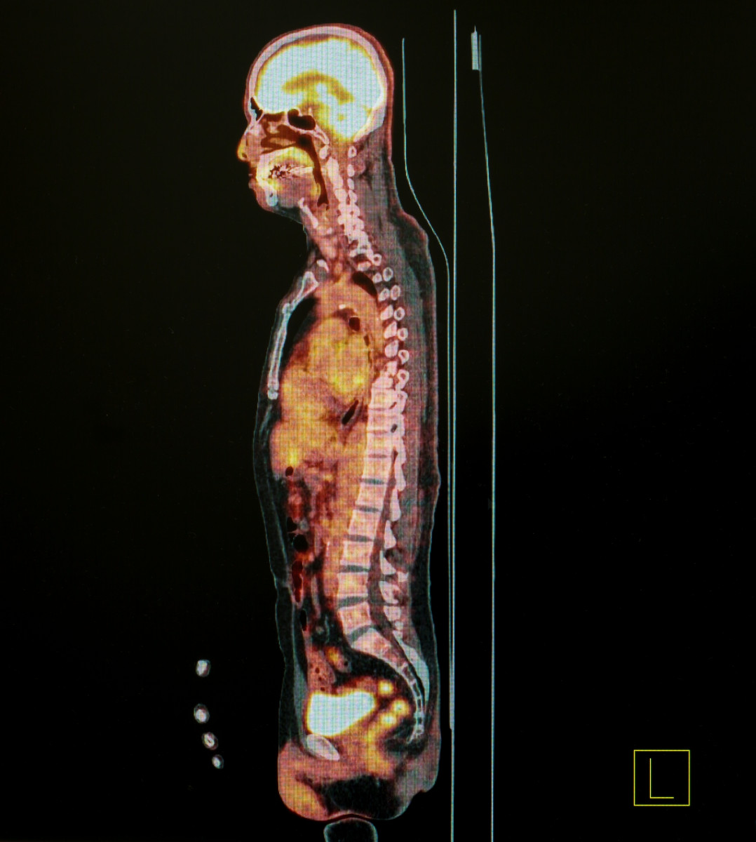


Researchers are developing a new positron emission tomography (PET) scanner capable of imaging the whole body that promises to significantly increase imaging sensitivity, advancing the technology for how diseases such as sarcoidosis are diagnosed and treated.
“It will offer the ability to detect throughout the whole body the location of focal pathologies, including cancer, infection, and inflammation [including sarcoidosis] at considerably lower levels of disease activity than is currently possible,” Terry Jones, study author and a clinical professor of diagnostic radiology at University of California, Davis, said in a press release.
The study “Total-Body PET: Maximizing Sensitivity to Create New Opportunities for Clinical Research and Patient Care” was published in the Journal of Nuclear Medicine.
PET is considered the most sensitive technique for assessing, in a non-invasive manner, key functions of the human body – from physiology to metabolism, and even glimpsing molecular pathways.
PET’s utility in the clinic, however, is still limited by low signal-to-noise ratio that affects the quality of the images and prolongs the time required for image acquisition. As a consequence, patients may undergo a longer exposure to radiation.
Now, a team of researchers at the University of California, Davis developed a PET scanner that promises to dramatically increase the sensitivity of PET. First, the new scanner can image the whole body at once, and its “sensitivity can be increased by a factor of about 40 for total-body imaging or a factor of about 4–5 for imaging a single organ such as the brain or heart,” researchers wrote.
Another advantage is that the new whole-body PET scan also can improve timing resolution significantly, which can lead to even further gains in sensitivity, and making the scans faster and reducing the amount of radiation exposure.
“Whole-body PET scans could be performed for a radiation dose roughly equivalent to that received from a round-trip transatlantic flight.” Jones said.
“It will also reduce the time taken to scan the whole body by at least a factor of 10, leading to scan times that could be less than one minute. This, for example, will make it far easier to scan infant and pediatric subjects without anesthesia or sedation,” Jones added.
Closer to First Test in the Clinic
The technology originally was developed for clinical research to allow scientists to understand how newly developed medicines move once in the body – a parameter known as pharmacokinetics – and accelerate the development of new therapeutic agents.
Now, UC Davis in collaboration with United Imaging Healthcare is building the first prototype total-body PET/CT (computed tomography) scanner, called Explorer, which is expected to revolutionize patients’ diagnosis and care.
“The applications of nuclear medicine will expand considerably across internal medicine at a rate not witnessed to date, and will become more evenly distributed across the age spectrum,” said Jones. “There will be a considerable stimulus/investment to develop new imaging biomarkers especially within immunology and endocrinology.”
While the cost of a whole-body PET scan is considerable, Jones highlighted that “as the impact of high-sensitivity, total-body PET scanning becomes apparent, this will provide a major stimulus to physicists, chemists, and engineers to develop lower-cost detectors for total-body surveillance.”
Jones said he believes that with this device we are witnessing the time of “nuclear medicine coming of age.”