Chest X-Ray for Sarcoidosis Screening in Uveitis Patients Requires More Studies
Written by |
An analysis of the usefulness of chest X-ray screening for sarcoidosis among patients with a certain type of eye inflammation (uveitis) failed to answer the question of whether it might be better to omit X-ray screening from initial evaluations in favor of more sensitive imaging techniques.
Researchers stated that this failure was a result of the incapacity to assess the accuracy of sarcoidosis detection using other methods; therefore future studies, using simultaneous screening with various methods, are needed.
The study, “Chest Radiographic Screening for Sarcoidosis in the Diagnosis of Patients with Active Uveitis,” was published in the journal Annals of the American Thoracic Society.
Since uveitis — an inflammation of the eye’s pigmented layer — can be linked to sarcoidosis, medical examination of patients with this type of eye inflammation includes chest X-rays. But researchers at Erasmus University and Erasmus Medical Center in the Netherlands noted that there is no published evidence demonstrating that this approach is effective.
The team analyzed the medical records of 200 patients who recently developed uveitis.
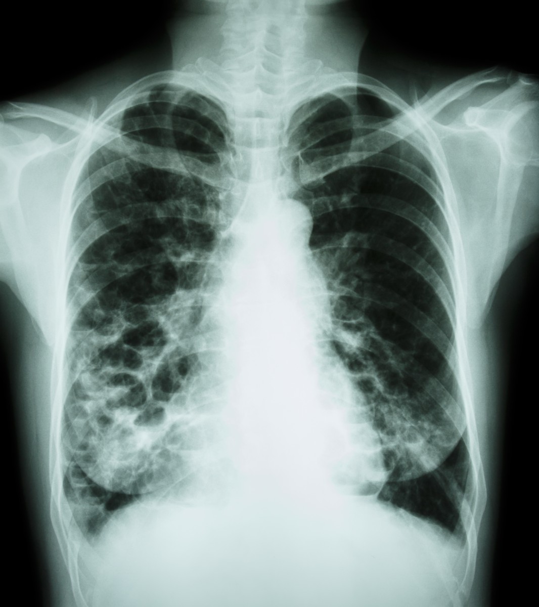
Abnormal chest X-rays were seen in 30 participants of the 200, amounting to 15%. Of these 30 cases, 43% showed changes that indicated the presence of sarcoidosis.
Ultimately, 22 patients (11%) were diagnosed with sarcoidosis through a biopsy. The initial chest X-rays had been abnormal for 14, or 64%, of these 22 patients.
Analyses showed that the interpretation of X-ray findings had a rather poor sensitivity, meaning that the X-rays were not particularly valuable for providing a positive diagnosis for the condition. Nevertheless, the ability to spot those without sarcoidosis was far better, being correct in 91% of the cases.
When combining chest X-rays with measures of angiotensin-converting-enzyme levels in the blood — often elevated in sarcoidosis — the sensitivity of the diagnosis was improved to 79%.
Most patients with sarcoidosis, confirmed by biopsy, also had a chest computed tomography (CT) scan that showed typical sarcoidosis changes. Researchers also noted that sarcoidosis, confirmed by biopsy, was more common in patients with panuveitis, a condition in which all the layers of the uvea are inflamed.
The team concluded that “a majority of the abnormal chest radiographs showed findings compatible [with] the diagnosis of sarcoidosis.” Still, the researchers emphasized they were unable to answer the question of whether X-ray screening is a necessary procedure within the initial evaluation of patients with uveitis, or if more sensitive imaging should be used in those who show signs of sarcoidosis.
To answer this question, a study using simultaneous imaging techniques based on X-rays, CT-scans, and somatostatin receptor scintigraphy tests would be needed.