
Higher Numbers of 2 Bacteria Species Found in Sarcoidosis Patients, Study Finds
Written by |
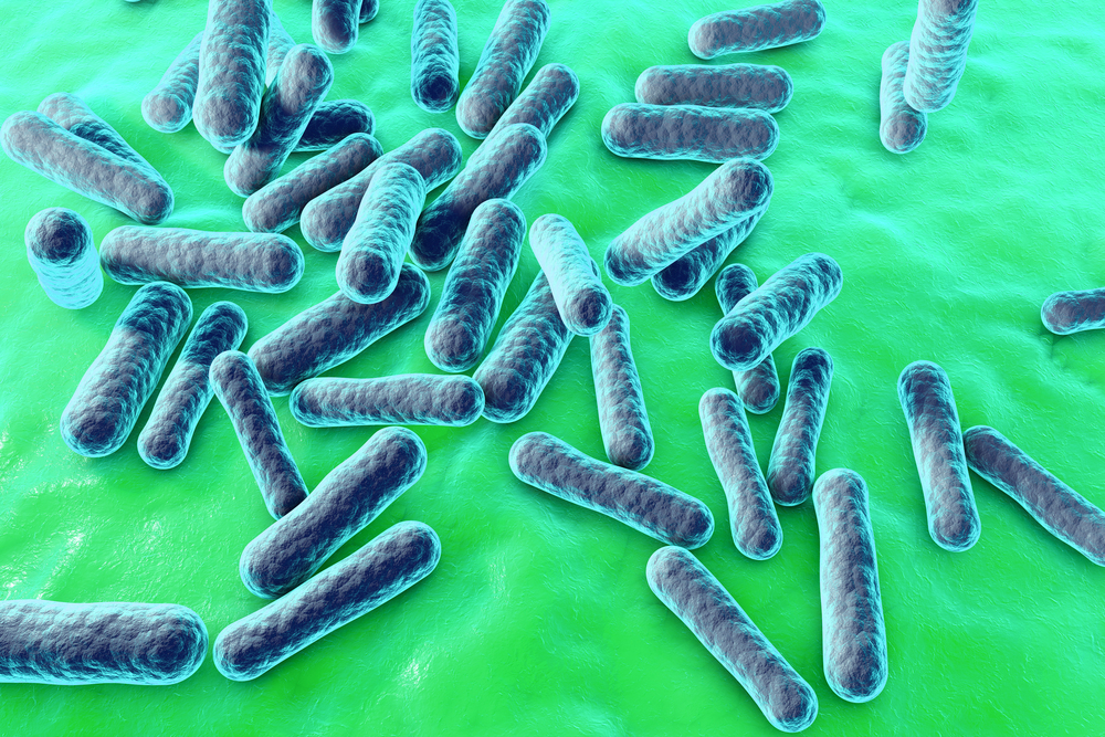
Researchers found higher numbers of two species of bacteria in the lungs of patients with sarcoidosis, and call for further studies to better understand their importance in the development of the disease.
The study, “Atopobium and Fusobacterium as novel candidates for sarcoidosis-associated microbiota,” was published in the European Respiratory Journal.
The study included 71 patients with sarcoidosis, ages 24 to 67, in a total of 39 men and 32 women. Also, 10 people participated as healthy controls (HC), and 15 patients with idiopathic pulmonary fibrosis (IPF) took part as an additional non-infectious patient group.
The many different types of bacteria present in the lungs of study participants were identified by DNA analysis of bronchoalveolar lavage (BAL) — a technique by which cells and fluid from bronchioles and lung alveoli are obtained.
Operational taxonomic units (OTUs), a way of identifying the species of bacteria present, differed between sarcoidosis, IPF, and the healthy control (HC) samples.
A bacterial species called Veillonella was more abundant in HC samples, and there were higher numbers of Streptococcus (OTU1) in HC and IPF samples. Unlike sarcoidosis samples, there were higher numbers of Atopobium (OTU41) and Fusobacterium (OTU16).
While Atopobium was present in 68% of the sarcoidosis samples, it was not detected in HC samples. Atopobium was found in IPF samples but less often and in lower numbers than in those with sarcoidosis.
A hallmark of sarcoidosis is the formation of granulomas, which are clusters of immune cells. Scadding’s criteria of stages are descriptions of the type of sarcoidosis in a patient, and are evaluated using chest X-rays.
Stage one sarcoidosis involves granulomas in the lymph nodes (small organs of the immune system found throughout the body). Stage two involves granulomas in lymph nodes and in the lungs. Stage three sarcoidosis indicates granulomas present in the lungs, but not in the lymph nodes.
Result showed that the numbers of Atopobium bacteria were significantly higher in sarcoidosis types one and two, compared to healthy controls and IPF samples. High numbers of Fusobacteria were found in types two and three sarcoidosis samples.
The team emphasizes that this study in no way indicates that Atopobia and Fusobacteria species cause sarcoidosis, but demonstrate a need for additional studies into the role they may play in disease progression.
“Taken together, our findings from BAL samples support the hypothesis of a sarcoidosis-specific microbiota, with Scadding types showing distinct bacterial patterns,” the researchers wrote, adding that “we found imbalances in the sarcoidosis microbiota, especially Atopobium spp. and Fusobacterium spp. associated with sarcoidosis-specific Scadding types.”
The team also highlighted “the need to further assess the role of lung microbiota for manifestation, progression and therapy of sarcoidosis, and its interplay with genetic factors.”


Steve Bruskiewicz
I also suffer from Sarcoidosis of the lung and would be interested in participating in studies if any are available in Wisconsin as I live in the Milwaukee area.