Low Lymphocytes Levels Can Help Diagnose Active Sarcoidosis
Written by |

An association has been found between active sarcoidosis, inflammation, and the presence of low levels of white blood cells, called lymphocytes, that are involved in immune defense.
This finding, by researchers at the University of Illinois Chicago (UIC), could be the first step toward the development of a new biomarker for sarcoidosis.
The results of the study, “Unsupervised Clustering Reveals Sarcoidosis Phenotypes Marked by a Reduction in Lymphocytes Relate to Increased Inflammatory Activity on 18FDG-PET/CT,” were recently published in the journal Frontiers in Medicine.
“Having a biomarker signature that indicates active inflammation could be very useful in helping guide treatment and for helping us understand the underlying mechanisms associated with the disease,” Nadera Sweiss, MD, the study’s senior author and chief of the department of rheumatology at the UIC College of Medicine, said in a press release.
Sarcoidosis can be characterized by periods of inactive disease and those of active disease, in which granulomas form in different tissues and organs. Granulomas are immune cell clusters that can affect how well organs work.
Being able to identify patients at risk of active disease is important to guide treatment.
Disease activity in sarcoidosis is associated with active inflammation. Therefore, the researchers analyzed blood markers of inflammation, as well as imaging data, and then assessed their possible relationship with disease activity.
In total, the medical records of 58 sarcoidosis patients — mostly African American (60.34%) and women (63.79%), and all between the ages of 30 and 69 — were analyzed; all were being followed at the Bernie Mac Sarcoidosis Translational Advanced Research (STAR) Center at UIC.
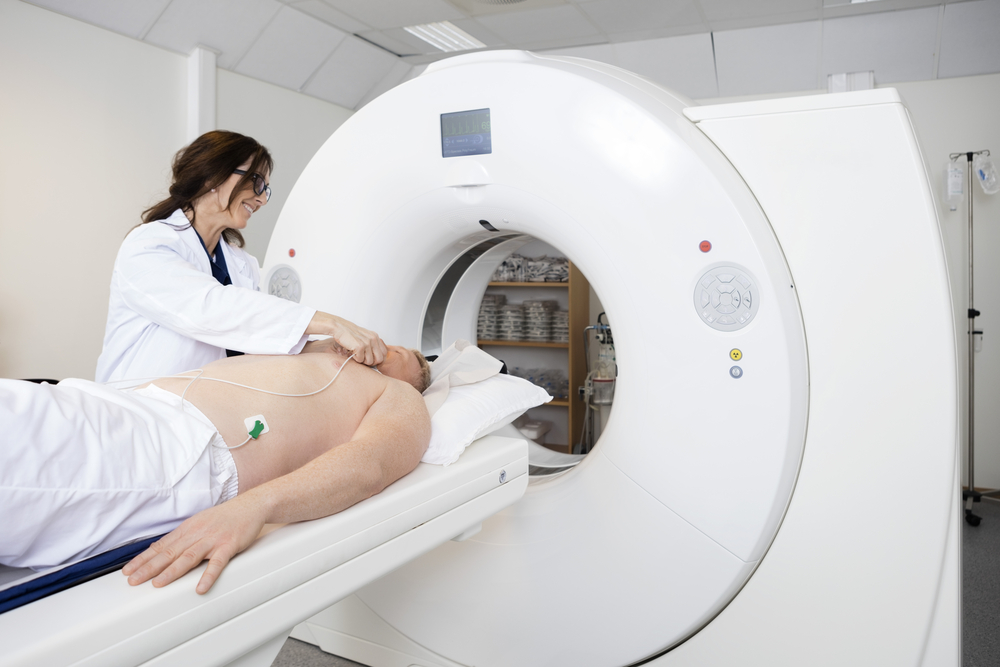
Skull to thigh PET/CT (positron emission tomography–computed tomography) scanning was done for all patients, in combination with a diagnostic tracer, called 18F‐fluorodeoxyglucose (FDG), to visualize areas of active inflammation.
The team used a statistical technique, called cluster analysis, to evaluate the relationship between parameters such as race, laboratory data, and the presence of inflammation.
The results showed that higher levels of inflammation on the PET/CT scans were linked with active sarcoidosis, as were low blood levels of lymphocytes.
“We found that active inflammation, as shown in PET/CT scans, was associated with active disease states,” said Christian Ascoli, MD, one of the study’s authors. “Lymphocyte levels were lower in patients with more inflammation, indicating that lymphocyte levels are also a marker of active disease.”
Interestingly, the team identified three groups, or clusters, of patients. One consisted of mainly African American patients with inactive disease, while another included African American individuals but with high levels of inflammatory markers and advanced pulmonary disease. A third cluster was comprised of mostly Caucasian patients with lower levels of lymphocytes and acute disease, and greater inflammation.
The second and third clusters had significantly higher levels of inflammation, as shown by PET/CT scans.
Further analysis showed that lymphocyte count levels could predict the degree of inflammatory activity detected by PET scans — with lower lymphocyte levels being associated with greater levels of inflammation.
Taken together, the team identified “three distinct sarcoidosis phenotypes with significant variation in race, disease chronicity, and inflammation,” they wrote.
The study was key in providing much-needed new information about sarcoidosis, the researchers said.
“Our finding represents another tool in our toolbox for guiding treatment of this very variable disease, and helping us learn more about its underlying biology,” Sweiss said.
According to the team, although further studies with larger patient groups are needed to confirm the findings, the data suggest that the levels of lymphocytes in the blood could be used together with PET scans to assist clinicians in deciding the best treatment approach for sarcoidosis patients.
“Our study opens new doors for research that will help implement new classification criteria for the diagnosis and treatment of sarcoidosis. Multicenter validation studies will help to determine if this classification scheme can be applied broadly as well as clarify the clinical and immunologic implications of these findings,” the team wrote.


