
Only 1 in 6 people get definite neurosarcoidosis diagnosis: Study
Findings confirm biopsy, while invasive, is still most valuable test
Written by |

Despite advances in testing, fewer than one of every six people evaluated for suspected neurosarcoidosis — sarcoidosis affecting the nervous system — received a definite diagnosis of the inflammatory disorder, according to a 30-year study in Sweden.
A confirmed neurosarcoidosis diagnosis in those cases was made only following a tissue biopsy of the central nervous system (CNS), comprised of the brain and spinal cord. Most of the other cases were classified as either probable or possible neurosarcoidosis, based on consistent signs and symptoms. Meanwhile, one-quarter of the individuals tested were confirmed to have another disease, based on CNS biopsy results.
These findings confirm a tissue biopsy as the most valuable test to resolve a suspected case of neurosarcoidosis. However, the researchers emphasized that the test’s invasive nature and associated health risks continues to limit its use.
“Since 2007, the employment of a structured laboratory and imaging approach and the increasing number of CNS biopsies have facilitated and improved the process of correct attribution in patients with presumptive neurosarcoidosis, especially in patients with isolated neurological lesions,” the researchers wrote, nonetheless noting the “lack of reliable biomarkers” for the disease.
The study, “A comprehensive diagnostic approach in suspected neurosarcoidosis,” was published in the journal Nature Scientific Reports.

Patients with possible neurosarcoidosis diagnosis spanned 3 decades
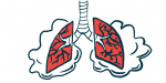
Sarcoidosis is a rare immune system disorder marked by the formation of tiny clusters of inflammatory white blood cells called granulomas. Such clusters can form in different parts of the body, most commonly in the lungs — when it’s referred to as pulmonary sarcoidosis — and disrupt organ and tissue function.
In up to 15% of cases, the disease affects the nervous system, and is then known as neurosarcoidosis. This rare form of sarcoidosis can be difficult to diagnose because it presents a wide range of non-specific symptoms and laboratory and MRI findings that also can be caused by other diseases.
That may include not only neurological and autoimmune disease such as multiple sclerosis, neuromyelitis optica spectrum disorder, lupus, and Sjögren syndrome, but also infections and certain types of cancer.
Given these complexities, a team of investigators at the Uppsala University Hospital, in Sweden, sought to describe their experience in diagnosing neurosarcoidosis. Their aim: to “highlight the ongoing challenges, focusing on the diagnostic workup.”
The researchers identified 90 people with a suspicion of neurosarcoidosis from 1990 to 2021 at their hospital. Most (60%) were classified as having possible neurosarcoidosis, while the remaining 40% were classified as having probable neurosarcoidosis.
However, a biopsy of CNS tissue revealed an alternative diagnosis in 24 patients (26.7%) initially classified as possible neurosarcoidosis. As such, the researchers then focused their analysis on the remaining 66 patients — 35 women and 31 men.
Their age at symptom onset ranged from 35-60 years, with a median age of 49. One-quarter were smokers, and nearly half were initially treated for a medical emergency.
Symptoms related to cranial neuropathies, or damage to nerves that control muscles and sensation in the head and neck, were the most common (38%). Of these, the optic nerve, which relays signs between the eyes and the brain, was most likely to be affected.
Other common early symptoms included movement problems (32%), headache (16%), and pituitary/hypothalamic dysfunction (12%). The pituitary gland and the hypothalamus are two brain regions commonly affected by neurosarcoidosis.
Non-specific symptoms also were noted, such vision and hearing problems, vertigo, nausea, chronic fever, seizures, confusion, cognitive impairment, hallucinations, and sexual dysfunction. The peripheral nervous system, comprising the nerves outside the brain and spinal cord, was affected in three patients.
A total of 36 patients (55%) had additional involvement outside the nervous system; of them, 29 had pulmonary sarcoidosis. Other affected tissues included the skin, salivary gland, lymph nodes, heart, joints, bone marrow, and the digestive system.
MRIs, biopsies used in making neurosarcoidosis diagnosis
A brain MRI was performed in 94% of patients, with spinal cord MRIs done in 52%.
One of the “most striking findings in MRI,” the researchers wrote, was the presence of isolated pituitary gland and hypothalamus damage in 17% of patients. That proportion was nearly twice as high as had been reported in previous studies (9%).
Other brain MRI findings included inflammation of the optic nerve in 15 patients (22%), while spinal cord images showed inflammation, tissue damage, and swelling.
Cerebrospinal fluid (CSF), the liquid surrounding the brain and spinal cord, was collected from 54 patients (82%), of whom 42 (78%) had abnormal test results. Most prominently, high levels of white blood cells were detected. These were not due to infections or other causes, however, which suggested a neuroinflammatory disease, the team noted.
During their initial analysis of medical records, the team found that 90 patients had been registered as either possible or probable for having neurosarcoidosis.
Besides the excluded 24 patients who were found to have a disease other than neurosarcoidosis, 14 others underwent CNS tissue biopsy, confirming a neurosarcoidosis diagnosis.
Most of the patients undergoing biopsy had been classified as having possible neurosarcoidosis, “based on their clinical presentations and the absence of systemic sarcoidosis,” the team wrote.
Importantly, the team added, a CNS biopsy was not performed in most of the patients classified as probable neurosarcoidosis “because of the presence of systemic sarcoidosis.”
Isolated CNS damage was found in the majority of patients with definite neurosarcoidosis.
“In reality, the most difficult scenario was investigating isolated neurosarcoidosis,” the researchers wrote.
The high number of patients with probable and possible diagnosis versus only 21% of cases with definite neurosarcoidosis reflect real world data.
Overall, 14 patients (21%) were classified as having definite neurosarcoidosis, while 32 were marked as probable sarcoidosis (49%) and 20 as possible neurosarcoidosis (30%).
“The high number of patients with probable and possible diagnosis versus only 21% of cases with definite neurosarcoidosis reflect real world data,” the team concluded.
Because CNS biopsy is invasive, “diagnosis relies on how well the clinician interprets the likelihood of differential diagnosis based on the patient´s history, clinical findings, results of neuroimaging, and CSF and [blood] analyses,” they added.


Leave a comment
Fill in the required fields to post. Your email address will not be published.