Study Supports Theory That Bacteria Causes Sarcoidosis, Researchers Say
Written by |
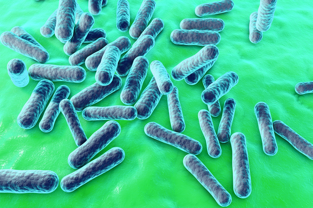
Researchers found bacteria of the type Propionibacterium acnes in a large proportion of tissue samples from patients with cardiac sarcoidosis, suggesting that the bacteria may cause the formation of granulomas (abnormal collections of inflammatory cells), a typical feature of sarcoidosis.
The work, published in the journal PLOS ONE, indicated that tests for the bacteria might be used to discriminate between cardiac sarcoidosis and other types of conditions affecting the heart.
The report is titled “Immunohistochemical identification of Propionibacterium acnes in granuloma and inflammatory cells of myocardial tissues obtained from cardiac sarcoidosis patients.”
The researchers at the Hokkaido University Graduate School of Medicine, together with colleagues at Tokyo Medical and Dental University, both in Japan, said their study also provides more solid proof that the bacteria causes the disease compared to earlier studies.
Researchers studied tissue from 26 patients with cardiac sarcoidosis along with samples from 15 myocarditis patients and 39 patients with other cardiomyopathies, or diseases affecting the heart muscle. Myocarditis is an inflammation of the heart muscle. The tissue samples were gathered from surgeries, tissue biopsies, and autopsies.
The team found granulomas in 62% of the sarcoidosis patients. None of the other patient samples had such granulomas.
Sarcoidosis patients had a similar proportion of high-grade inflammatory lesions as patient with myocarditis — 62% and 67%, respectively. Those with other heart muscle conditions had much lower numbers of such lesions — they were found in only 3% of the samples.
Researchers also found that 63% of granulomas from cardiac sarcoidosis patients tested positive for P. acnes. The bacteria were found in 63% of high-grade inflammatory lesions from sarcoidosis patients and in 73% of inflammatory lesions with a lower grade of inflammation.
Again, researchers could not detect the bacteria in inflammatory lesions from samples of patients with the other analyzed heart conditions.
The team found no difference in the presence of bacteria between patients with specific symptoms, including symptoms not related to the heart.
Researchers underscored that the presence of P. acnes bacteria in granulomas and lesions in the heart — which, under normal conditions, does not contain bacteria — is a stronger proof of the bacteria’s involvement in sarcoidosis than studies in lung and lymph nodes.
Although several earlier studies have found P. acnes in sarcoidosis granulomas in the lung and lymph nodes, these organs may also contain P. acnes in the absence of disease.
The team, therefore, believes that P. acnes might be the factor driving sarcoidosis in many patients. The samples in which the bacteria were not found might instead be infected with other types of bacteria such as Propionibacterium granulosum and Mycobacterium tuberculosis, the researchers hypothesized.
Researchers also suggested that the finding of P. acnes in inflammatory lesions and granulomas might be used to aid in the diagnosis of cardiac sarcoidosis.