
1st cardiac sarcoidosis mouse model may help develop new therapies
Researchers use model to identify protein called mTORC1 as potential target
Written by |

Researchers in Austria and the U.K. have created the first mouse model that mimics the human disease of cardiac sarcoidosis.
“To our best knowledge this is the first animal model of cardiac sarcoidosis that recapitulates major … hallmarks of human disease,” scientists wrote, adding that it is expected to “allow for mechanistic insights into the molecular events underlying the disease and suggest novel therapeutic targets.”
Indeed, studies in the model allowed researchers to identify a protein called mTORC1 as a potential target in cardiac sarcoidosis and to support clinical testing of suppressors of mTOR proteins, including some already used for other conditions, in this patient population.
The team described the model and associated findings in the study “An mTORC1‐Dependent Mouse Model for Cardiac Sarcoidosis,” which was published in the Journal of the American Heart Association.

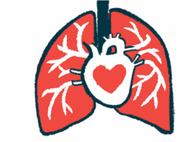
Cardiac sarcoidosis is severe complication when granulomas affect the heart
Sarcoidosis is characterized by clumps of inflammatory immune cells called granulomas, which can form in various organs in the body causing damage and scarring (fibrosis). Cardiac sarcoidosis, when these granulomas affect the heart, is one of the most severe complications, though it can be hard to accurately diagnose and treat.
Mouse models are a key research tool for understanding how diseases work and testing potential treatments, but to date, there hasn’t been an established mouse model of cardiac sarcoidosis.
Here, a team of researchers at Medical University of Vienna, in Austria, and St. George’s, University of London, in the U.K., developed such a model by genetically engineering mice so that a certain subset of myeloid cells lack the TSC2 gene.
Myeloid cells are a class of immune cells, including macrophages, that are centrally involved in granuloma formation. The TSC2 gene normally acts to limit the activity of the mammalian target of rapamycin complex 1, or mTORC1, a protein involved in the regulation of cell metabolism and immune response.
Previous studies by the same team had shown that genetic deletion of TSC2 in certain myeloid lineage cells “led to spontaneous development of pulmonary sarcoid‐like granulomas in mice,” the researchers wrote.
Without TSC2, mTORC1 becomes abnormally active, leading to uncontrolled growth of macrophages and recruitment of other cells, including fibroblasts, that eventually lead to granulomas and fibrosis.
The researchers found that their mouse model, modified to have overactive mTORC1 in certain myeloid cells, spontaneously developed granulomas in the heart.
“We have now discovered that the permanent activation of the protein mTORC1 directly in macrophages is sufficient for granulomas to develop spontaneously in the heart,” Clarice Lim, PhD, one of the study’s authors and a postdoctoral researcher at the Medical University of Vienna, said in a university news story.
To our best knowledge this is the first animal model of cardiac sarcoidosis that recapitulates major … hallmarks of human disease.
Heart abnormalities in mice comparable to what’s seen in patients
Further analyses showed the mice developed more and more granulomas in the heart, progressively worsening heart fibrosis, and showed reduced heart function and heartbeat abnormalities that were overall comparable to what’s commonly seen in people with cardiac sarcoidosis.
Of particular note, data indicated that this model has abnormalities in the connections between heart cells known as gap junctions. Problems with these connections can set the stage for irregular heart rhythms, which are a common cause of health problems in people with cardiac sarcoidosis.
The researchers demonstrated that most of these abnormalities could be reversed by treating the mice with everolimus, a medication approved for several types of cancer that works by blocking the mTORC1 protein.
Treatment with another compound called Bay11‐7082, which suppresses an immune protein called NF-kB, did not show beneficial effects in the mouse model.
Importantly, the scientists also detected mTORC1 overactivation in heart samples from seven out of nine people who died due to cardiac sarcoidosis.
Collectively, these data indicate that mTORC1 activation may play a role in driving cardiac sarcoidosis in people and that therapies blocking this protein may be useful in treating this condition.
“As everolimus treatment clears inflammatory infiltrates and improves cardiac function and fibrosis in this model of sarcoidosis, we propose the use of mTOR inhibitors in future clinical trials to test for efficacy” in cardiac sarcoidosis, the scientists wrote.

Mouse model also showed granulomas in the lungs
Notably, besides cardiac sarcoidosis, the mouse model also showed granulomas in the lungs, a form called pulmonary sarcoidosis.
Using this model, the Medical University of Vienna team, together with colleagues at Vanderbilt University School of Medicine, in Texas, was able to identify another therapeutic target for sarcoidosis: an immune-related protein called SHP2.
Results were published in Science Translational Medicine in a study, titled “SHP2 promotes sarcoidosis severity by inhibiting SKP2-targeted ubiquitination of TBET in CD8+ T cells.”
The team showed that suppressing SHP2 reduced the formation of large granulomas and numbers of macrophages in the lungs of the mouse model.
However, this approach “only had a therapeutic benefit in about 50% of the animals in our model, but interestingly not in the others,” Lim said.
Further analyses showed that blocking SHP2 was only beneficial in mice who had high levels of this protein in a particular class of immune cells called CD8-positive T-cells.
The scientists found that CD8-positive T-cells producing high SHP2 levels would secrete signaling molecules that recruited other immune cells to the granuloma, driving disease progression.
Blocking SHP2 could interrupt this process, and the researchers showed it had similar effects in immune cells collected from people with progressive pulmonary sarcoidosis and end-stage disease.
“These studies provided new mechanistic insights into the [biological function of SHP2] and identified SHP2 inhibition as a potential therapeutic intervention against severe sarcoidosis,” the scientists wrote.


Leave a comment
Fill in the required fields to post. Your email address will not be published.